Congenital Urethral Polyps: A Rare Cause of Bladder Outlet Obstruction in Children-Juniper Publishers
Juniper Publishers-Journal of Pediatrics
Abstract
Polypoid mass in the prostatic urethra is uncommon
but potentially represents a wide spectrum of different entities,
ranging from congenital malformations, benign polyps, premalignant
disorders to various malignancies. Congenital urethral polyps are rare
entity in infancy. They are congenital benign tumors. Most patients
present with acute retention, hematuria or intermittent bladder outlet
obstruction. The treatmentof choice is transurethral endoscopic
ortransvesical resection. Histological study confirms the diagnosis and
theprognosis is excellent. We report a case of an 18 month's old boy who
presented with acute urinary retention. There was history of dribbling
of urine and excessive crying during micturition. Imaging and endoscopic
studies confirmed the presence of a polypoid pedunculated lesion at the
bladder base arising from posterior urethra. It was excised by
cystostomy because of an unsuccessful cystoscopic removal attempt.
Although this is considered a benign lesion with no previous reports of
recurrence or malignant behavior, it produces dramatic urinary symptoms
in the pediatric population with a wide differential diagnosis. Imaging
and endoscopic findings may suggest a malignancy and are not sufficient
to render a precise diagnosis, which can only be made by pathologic
examination.
Keywords: Polyps; Urethra; Hematuria; Congenital; Urinary retention
Introduction
Congenital urethral polyps cause a variety of
symptoms in children. John Hunter is credited with the first documented
case of urethral polyp, and Thompson reported the first case in a
patient. Congenital urethral polyps are rare with only sporadic reports
of a small series of cases [1].
Posterior urethral polyps in children are uncommon, and anterior
urethral polyps are even rarer. Nevertheless, anterior urethral polyps
are exceptionally reported in the literature [2-5].
These polyps are congenital and occur usually in boys; the average age
of presentation is 5.2 years. Polyps in boys mostly arise from the
posterior urethra, are usually proximal to the membranous urethra, and
present usually as a single tumor, only rarely as multiple separate
masses. This benign pathology is supposed to represent a developmental
error in the invagination process of the submucosal, glandular portion
of the inner zone of the prostate gland [6].
These Congenital urethral polyps are the most common benign mesodermal
tumors of the urinary tract. Such lesions can also be called prostatic
urethral polyps (in boys), fibroepithelial polyps of the urethra, or
benign urethral polyps. Congenital urethral polyps more frequently occur
in males and in the posterior urethra; this entity is very rare in
females. Congenital urethral polyps should not be confused with the
polyps of botryoid sarcoma [7].
The embryologic basis for urethral polyps is not clear; however, it is
possible that they may arise from mesonephric remnants [8].
These lesions are benign, pedunculated tumors that arise in the region
of the verumontanum in males or mid-urethra in females. In early
infancy, they usually cause urethral obstruction. However, in older
boys, the main presenting features include hesitancy, diminished stream,
incomplete emptying, urinary retention, and sudden painful interruption
of urinary stream, dysuria, hematuria, UTI, VUR, and enuresis.
Case Report
An 18-month-old boy presented with complaint of
urinary retention and dribbling of urine for 3 days. Clinical
examination revealed no abnormalities. Laboratory assessments were
unremarkable, except for 4-5 red blood cells on urine analysis.
Cystourethrography (VCUG) revealed normal findings and ultrasonography
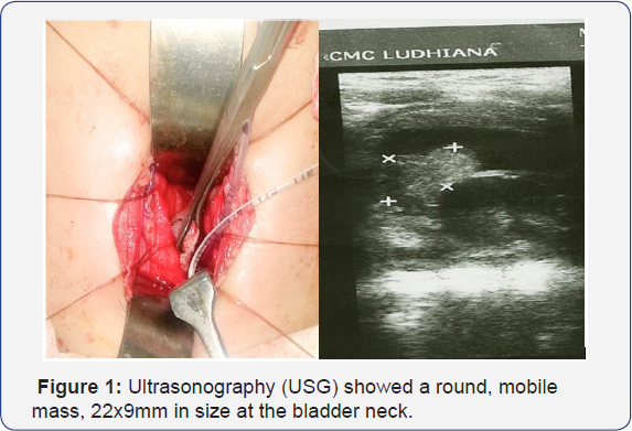
(USG) showed a round, mobile mass, 22x9mm in size at the bladder neck.
Cystourethroscopy confirmed the presence of a posterior urethral polyp
extending from posterior urethra into the bladder. An attempt to remove
it endoscopically was unsuccessful, because the polyp had a smooth
surface, a tense wall and was floating in the bladder Therefore, it was
excised by cystostomy. Pathologic gross findings consisted of polypoidal
greyish white soft tissue measuring 1.5x1x1cm. Micro- section shows a
polyp covered by transitional epithelium which is locally ulcerative,
other areas the epithelium is mildly hyperplastic. The stroma is
composed of vascular fibro-connective tissue and shows a dense
inflammatory infiltrate comprising of neutrophils, lymphocytes,
eosinophils and histiocytes (Figure 1).

Discussion
Congenital polyps of the male urethra are pedunculated lesions that arise as a defective protrusion of the urethral wall [1]. They occur more often in children and in patients present with bladder outflow obstructions [9].
The investigation of choice is urethrocystoscopy. An ultrasound of the
urinary bladder and posterior urethra may reveal a polyp.
Histologically, the polyps are composed of vessels, muscle and, less
frequently, nerves and glands covered by urothelium. There is some
controversy over their embryological origins [7-9]. Downs postulated that the polyps resulted from a defective protrusion of the urethral valve [1] . Kuppusami & Moors [10]
suggest that metaplastic epithelial changes had occurred secondary to
the estrogen released during gestation. They found similarities between
the urethral polyp and verumontanum histology, which consisted of smooth
muscle and small glands lined by transitional cell epithelium [10]. Stephens [11] postulated that the polyp is most likely a congenital lesion. Barrie & Simms [12]
believe that the polyp is a response to urinary tract infections;
however, most such inflammatory polyps do not occur in children [11].
The absence of glandular elements within the fibrous polyps of most
patients distinguishes them from the verumontanum. These polyps arise
from the vestiges of Muller's tubercle and then fail to regress [1].
A precise history, physical examination, and uroflowmetry patterns in
toilet-trained children can strongly suggest urethral polyps in
children. Urinary tract ultrasonography and micturition
cystourethrography are the most important examinations for this
diagnosis; however, they are not always successfully diagnostic. The
final diagnosis must be confirmed by direct video urethrocystoscopy with
minimal irrigation flow. Transurethral resection of the urethral polyps
is the standard firstline of management [13].
However, when the polyp length is larger than 3cm, with diameter of 1cm
or more, it becomes displaced into the bladder, and open transvesical
removal may be an acceptable alternative.
Conclusion
In all children who present with sudden interruptions
of their urinary flow and no obvious bladder or urethral stones,
urethral polyps must also be kept in mind as a differential diagnosis.
The final and definite diagnosis of polyps is made based on the
appearance on urethrocystoscopy. The endoscopic resection of the polyp
is the treatment of choice; however, transvesical resection is a safe
option in large bladder polyps.
For more articles in Academic Journal of
Pediatrics & Neonatology please click on:
https://juniperpublishers.com/ajpn/index.php
https://juniperpublishers.com/ajpn/index.php
Comments
Post a Comment