Congenital Prepubic Sinus (Type 2 Stephens Variant of Epispadiac Dorsal Urethral Duplication): An Uncommon Anomaly-Juniper Publishers
Juniper Publishers-Journal of Pediatrics
Abstract
Variants of congenital prepubic sinus have been
reported rarely, and because the anatomic features often differ from
each other, a consensus concerning the embryology and classification was
not achieved yet. Various names including congenital prepubic sinus,
sub pubic fistula, prepubic dermoid sinus, and suprapubic dermoid sinus,
were used to identify these lesions, and, among the classifications
available, none seems to clearly describe this entity. We present a
2-year old boy with a case of epispadiac variant of urethral duplication
in which the duplicated urethra presented as a prepubic sinus. We
report this uncommon anomaly and review the scattered published reports
to improve the global understanding of this uncommon congenital lesion.
Keywords: Urethral duplication; Epispadias; Congenital prepubic sinus; Sinus tract Introduction
Congenital prepubic sinus (CPS) is a rare anomaly of
uncertain etiology. The sinus usually presents as a small tract,
commencing on the skin overlying the penis or prepubic area, and
extending toward the anterior bladder wall or umbilicus [1,2].
The anatomic and pathologic features of this disorder have been
documented, but controversies over its embryologic basis are ongoing.
Case Report
A 2-year-old male child was presented with congenital
opening over the dorsal surface of the penis. He was asymptomatic
except for occasional clear discharge from the opening. Child was
passing urine through the normal urethral opening. Local examination
revealed a deformed penis with ventrally hooded prepuce and 8mm midline
epispadiac opening over the dorsal surface of the penis. A number 8
infant feeding tube could be easily passed in the opening for a distance
of 3cm and could be felt going under the pubic symphysis (Figure 1a & 1b).
Fistulography was showing blind ending tract around 2.5cm long going up
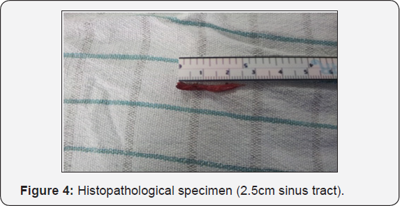
towards the prevesical space without any communication with bladder (Figure 2a & 2b). At operation, the tract was dissected and was found to be going under the pubic symphysis (Figure 3a & 3b).
The tract was traced up to prevesical space, transfixed, ligated and
divided. Specimen (2.5cm sinus tract) was sent for histopathology which
showed tract lined by transitional epithelium and focalinflammation (Figure 4).


Discussion
Congenital prepubic sinus is a tract originating in
the skin overlying the symphysis pubis, superior to the base of the
penis or clitoris, and extending to, but not communicating with, the
anterior bladder wall [3].
There are four generally proposed theories for the etiology of
Congenital prepubic sinus: First anomaly of abdominal wall closure [4]; and second urethral developmental anomaly, a variant of dorsal urethral duplication [1,2,5-8].
The third theory is that it is a congenital fistula of the primitive
urogenital sinus, with three anatomic subtypes depending on the
direction of the sinus tract: high, toward the urachal remnant; middle,
toward the bladder; and low, toward the prostatic urethra [9]. Fourth theory suggests that it is a remnant of the cloaca [10]. Of all these theories, reports supporting the anomaly of dorsal urethral duplication predominate [1,3,5-8,11].
As these theories cannot explain all the varied presentations of CPS,
Tsukamoto et al. postulated recently that CPS may be caused by a
residual cloaca membrane and umbilicophallic groove, and that the depth
may determine the position of the end of the sinus tract [12]. However, Huang et al. [1]
used an immunohistochemical staining technique of the excised sinus to
reinforce the theory of dorsal urethral duplication in a report of five
patients with congenital prepubic sinus, when they found the presence of
transitional epithelium in the proximal part of the sinus with
surrounding smooth muscle bundles [1]. Balster et al. [11]
in 2003 also supported this assumption with an immunohistochemical
study on the excised sinus tract of a 2-year-old boy with a skin fistula
on the dorsal side of the penis [11].
Urethral duplication remains a rare and confusing problem, more so when
it presents as a prepubic sinus. Urethral duplication does not
represent a uniform entity making it difficult to find an unequivocal
and comprehensive classification. Stephens described three types of
dorsal urethral duplication according to the anatomy [13].
Type 1 is a complete or incomplete channel that runs parallel to the
normal urethra from the glans to the bladder, which may join the urethra
or ends blindly. Type 2 is an epispadiac type of channel from the
dorsum of the penis to the bladder or one that joins the urethra at some
point. Type 3 is a dermoid sinus that simulates an accessory urethra
but tracks from the base of the penis in front of the pelvic urethra and
bladder behind the pubic symphysis to or towards the umbilicus. We
found only 7 reports of 9 cases of epispadiac variant of dorsal urethral
duplication in the English literature (Table 1) (6,8,14-18).
The similarity of the anatomy of our case to the type 2 variant of the
Stephens classification favours the theory that Congenital prepubic
sinus is a variant of dorsal urethral duplication. The presence of
transitional epithelium in the lining of the sinus in this patient
reinforced this theory. Although the tract ended blindly toward the
anterior bladder wall, the presence of dorsal chordee, a ventrally
hooded prepuce as well as penile torsion supports an epispadiac variant.

Patient with Congenital prepubic sinus should be
thoroughly evaluated because of its variable presentation, come to
medical attention because of the opening or because of persistent
discharge. Diagnosis is mainly clinical but imaging techniques such as
micturating cystourethrogram and sinogram could help to outline the
direction of the tract, and whether it is blind ending or communicating
with the urinary tract. Treatment is individualized depending on the
anatomy and severity of the anomaly, and usually consists of excision of
the non dominant urethra or sinus tract, usually the dorsal one.
Excision is usually curative.
For more articles in Academic Journal of
Pediatrics & Neonatology please click on:
https://juniperpublishers.com/ajpn/index.php
https://juniperpublishers.com/ajpn/index.php
Comments
Post a Comment