Congenital-Infantile Fibro sarcoma of Hand – A Case Report-Juniper Publishers
Juniper Publishers-Journal of Pediatrics
Abstract
Congenital infantile fibro sarcoma is a rare
malignant tumor of the soft tissue in the infant primarily developed in
the soft tissue of distal extremities and occasionally in unusual
location such as the lungs and retro peritoneum. It occurs mainly in
children below the age of 5 years. About 250 cases have been reported in
the literature so far, very few of them in newborns. Its clinical
course differs from conventional fibro sarcoma, the prognosis is
relatively good compared to adult forms and the 5 year survival rate can
be more than 80%. We report a case of congenital infantile fibro
sarcoma of the hand treated successfully by below elbow amputation. The
child is now 5 years old with no recurrence.
Keywords: Fibro Sarcoma; Congenital; InfantileIntroduction
Congenital infantile fibro sarcoma is an uncommon
soft tissue malignancy. It usually affects the extremities [1]. Few
cases involving the hand were reported and a limited number were
presents during neonatal period. It represents less than 1% of all
childhood malignancy, but it is the most common soft tissue sarcoma in
less than 1 year of age. Clinically there may be rapid growth of the
tumor so the initial clinical diagnosis is often erroneous [2]. Although
histological similar to fibro sarcomas occurring in adults,
congenital-infantile fibro sarcomas differs in their clinical behavior,
metastasis is rare and local recurrence is common. The prognosis is
good, much better than that for histological similar fibro sarcoma seen
in adults. In one series the 5 year survival rate for
congenital-infantile fibro sarcoma was more than 80%, which is in sharp
contrast to fibro sarcoma in adults, where 5 year survival rate is only
35% [3]. We report a case of congenital infantile fibro sarcoma
involving the left hand in a 25 days old male infant, managed by forearm
amputation and followed up for 5 years without recurrence or
metastasis.
Case Report
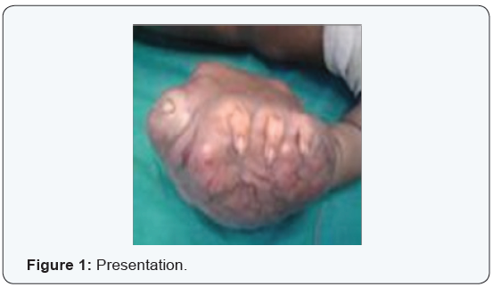
A 25 days old baby present with painless swelling of
the left hand. Parents had noticed a nodule on the palm of the left hand
since birth, which enlarged rapidly. The child was treated with
different antibiotic and homeopathic medicine locally. Because of rapid
enlargement of the swelling, the child was referred to our hospital when
he was about 1 month old. On physical examination he was a normal
healthy baby of 3 kg weight and a mass in the left hand which was poorly
circumcised, measuring about 8cm x 8cm, firm in consistency, skin was
tense and shiny. Other physical examination reveal no abnormality.
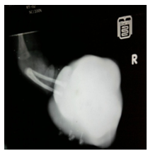
Plain x-ray of the left hand showed a huge soft
tissue mass. The laboratory tests, x-ray chest, abdominal
Ultrasonography was normal. Fine needle aspiration cytology (FNAC) of
the mass was done. It showed malignant spindle cell tumor. The size of
tumor was rapidly increasing doubling its size within 2 weeks. Open
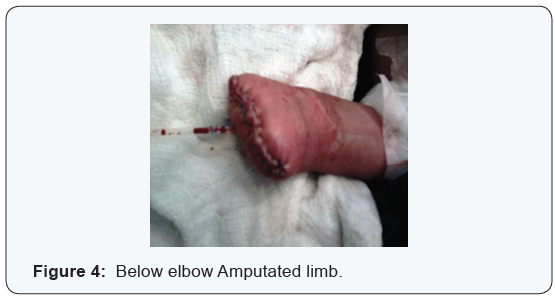
biopsy was done which suggested soft tissue sarcoma. The management was
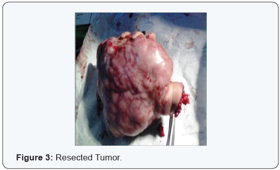
an below elbow amputation and his to pathological examination confirmed
the diagnosis. Gross pathological specimen showed skin with underlying
tumor tissue which infiltrating fat, skeletal muscle, bone and
neuro-vascular bundle. It was not involving the respected margins. Cut
surface was gray white with area of necrosis. Microscopic examination
confirmed features of infantile fibro sarcoma with Herring bone pattern.
Postoperatively the baby was stable. The child was followed up for 5
years of age without evidence of recurrence or metastasis.
Discussion
The congenital infantile fibro sarcoma is a rare soft
tissue malignant tumor. Fibro sarcoma is primarily an adult malignancy
and histological similar to those seen in children. The congenital
infantile fibro sarcoma usually occurs at birth or in the neonatal
period, mostly presenting within the first six months of life [4]. No
significant sex predilection has been found [3].
These tumors originate from the primitive mesenchymal
tissue. There is no evidence of increased familial incidence but a
recent study showed that all patients with congenital infantile fibro
sarcoma under the age of two years exhibited some type of chro mosomal
gain. Most had trilogies of chromosome 8, 11, 17 and/or
20 and an abnormal karyotype 48 xy, 11 and 20 [5,6].
Clinically, they present with a rapidly enlarging swelling in the
soft tissue most commonly in the extremities (71%) but may present
axial locations (29%) as well. When the limbs are involved, the
tumor tends to affect the leg more than the arm and the distal extremity
more than the proximal one [1]. A limited number however
developed primarily in the bones [7]. Therefore the initial clinical
diagnosis is often erroneous [2]. The diagnosis is sometimes
made base on a fast growing soft tissue swelling. At times it is
discovered late before one year [8]. On plain x-ray showed soft tissue
swelling that may obliterate normal fat planes and deform or
destroy the adjacent bone. CT scan is useful in demonstrating the
extent of the tumor and the amount of bony involvement, where as
MRI is particularly useful in showing the disruption of the soft tissue
planes. The MRI findings of congenital infantile fibro sarcoma
usually include a mixed cystic and solid tumor of heterogeneous
density which typically has in homogenous enhancement [6]. Although
the imaging findings are non specific, congenital infantile
fibro sarcoma should also be considered in cases of a soft tissue
mass in infants. A biopsy should be done to avoid delayed and incorrect
management [9].
Antenatal and postnatal ultrasound usually shows a poorly
circumscribed, heterogeneous and vascularize soft tissue mass,
which grows very rapidly and cause deformity of the anatomical
region involved, sometimes it causes polyhydramnios. Rest of the
fetus is usually normal. The diagnosis is done by the histological
examination having an plastic spindle shaped cells arranged in a
herring bone pattern, however, some of the striking features are
uniform, well oriented fibroblast, scattered round cells and chronic
inflammatory cells like lymphocytes. Multinucleated giant cells
are rare. Mitotic figures are common features. Rich vascular areas
may be seen [10].
Although histological similar to fibro sarcoma in adults, congenital-
infantile fibro sarcoma differs in its clinical behavior compared
with the adult type. Metastases are rare, however, local recurrence
is common. The prognosis for congenital-infantile fibro
sarcoma is extremely good with a 5 year survival rate of 84% in
series of 53 cases [2]. Treatment is wide local excision or amputation.
Chemotherapy and radiotherapy are reserved for unrespectable
tumor and for recurrence or metastasis. Amputation is indicated
only in the cases of local recurrence, giant tumor involving
bone and neurovascular structures [8].
Conclusion
In the case of our child the presence of diffuse soft
tissue involvement
of the hand and the bony lesions incite us to do a biopsy
which revealed a well differentiated congenital fibro sarcoma. The
extent of the tumor growth made us to perform an below elbow
amputation of the hand. That gave us a good result because no
metastasis or local recurrence were noted after 3 years however
follow up should be longer to look for these evolution aspects [11].
There are high metastatic and mortality rate for axial lesions,
indicating a more aggressive behaviors. The relatively benign course
of such tumors may be due to a significantly lower proliferative
index coupled with enhanced apoptosis. The favorable clinical
course and biological features of congenital-infantile fibro sarcomas
have raised a question about its nomenclature as sarcoma
rather than a borderline tumor [5,10].
Summary
In summary, although congenital-infantile fibro sarcoma is a
rare soft tissue tumor in children, it should be included in the differential
diagnosis of a large extremity mass presenting at birth
because the prognosis is much better than adult type. CT provides
the information about bony involvement but shows poor demarcation
of the extent of the tumor. On the other hand, MRI is particularly
useful in delineating the extent of the tumor. It also provides
multilane views so that diagnosis and planning for treatment can
be reinforced (Figures 1-4).


Comments
Post a Comment